Monday 22 June 2009

Mitral Stenosis


Mitral stenosis (mitral valve stenosis) is a narrowing of the mitral valve opening that increases resistance to blood flow from the left atrium to the left ventricle.

*
Mitral stenosis usually results from rheumatic fever, but infants can be born with the condition.
*
Mitral stenosis does not usually cause symptoms unless it is severe.
*
Doctors make the diagnosis after hearing a characteristic heart murmur through a stethoscope placed over the heart.
*
Treatment includes use of diuretics and beta-blockers or calcium channel blockers.

Animation

Understanding Mitral Valve Stenosis

In mitral stenosis, blood flow through the narrowed valve opening is reduced. As a result, the volume and pressure of blood in the left atrium increases, and the left atrium enlarges. The enlarged left atrium often beats rapidly in an irregular pattern (a disorder called atrial fibrillation). As a result, the heart's pumping efficiency is reduced. If mitral stenosis is severe, pressure increases in the blood vessels of the lungs, resulting in heart failure with fluid accumulation in the lungs and a low level of oxygen in the blood. If a woman with severe mitral stenosis becomes pregnant, heart failure may develop rapidly.

Cause

Mitral stenosis almost always results from rheumatic fever, a childhood illness that sometimes occurs after untreated strep throat or scarlet fever (see Bacterial Infections: Rheumatic Fever). Rheumatic fever is now rare in North America, Australasia, and Western Europe because antibiotics are widely used to treat infection. Thus, in these regions, mitral stenosis occurs mostly in older people who had rheumatic fever and who did not have the benefit of antibiotics during their youth or in people who have moved from regions where antibiotics are not widely used. In such regions, rheumatic fever is common, and it leads to mitral stenosis in adults, teenagers, and sometimes even children. Typically, when rheumatic fever is the cause of mitral stenosis, the mitral valve cusps are partially fused together.

Mitral stenosis can rarely be present at birth (congenital). Infants born with the disorder rarely live beyond age 2, unless they have surgery.

Three rare conditions unrelated to mitral stenosis can produce the same effects as the stenosis. They include a myxoma (a noncancerous tumor in the left atrium), cor triatriatum (a rare developmental abnormality in which a membrane goes across the left atrium), and pulmonary veno-occlusive disease (a narrowing of the veins that lead from the lungs into the left atrium).

Symptoms and Diagnosis

Mild mitral stenosis does not usually cause symptoms. Some people with more severe mitral stenosis have atrial fibrillation or heart failure. People with atrial fibrillation may feel palpitations (awareness of heartbeats). People with heart failure become easily fatigued and short of breath. Shortness of breath may occur only during physical activity at first, but later, it may occur even during rest. Some people can breathe comfortably only when they are propped up with pillows or sitting upright. Those people with a low level of oxygen in the blood and high blood pressure in the lungs may have a plum-colored flush in the cheeks (called mitral facies). People may cough up blood (hemoptysis) if the high pressure causes a vein or capillaries in the lungs to burst. The resulting bleeding into the lungs is usually slight, but if hemoptysis occurs, the person should be evaluated by a doctor promptly because hemoptysis indicates severe mitral stenosis or another serious problem.

With a stethoscope, doctors may hear the characteristic heart murmur as blood tries to pass through the narrowed valve opening from the left atrium into the left ventricle. Unlike a normal valve, which opens silently, the abnormal valve often makes a snapping sound as it opens to allow blood into the left ventricle. The diagnosis is usually confirmed by electrocardiography (ECG), a chest x-ray showing an enlarged atrium, and echocardiography, which uses ultrasound waves to produce an image of the narrowed valve and the blood passing through it.

Prevention and Treatment

Mitral stenosis will not occur if rheumatic fever is prevented by promptly treating strep throat with antibiotics. Treatment includes use of diuretics and beta-blockers or calcium channel blockers. Diuretics, which increase urine formation, can reduce blood pressure in the lungs by reducing the volume of circulating blood. Beta-blockers, digoxin Some Trade Names
LANOXIN
, and calcium channel blockers help control heart rhythms. Anticoagulants may be needed to prevent clot formation in people with atrial fibrillation.

If drug therapy does not reduce the symptoms satisfactorily, the valve may be repaired or replaced. Sometimes the valve can be stretched open using a procedure called balloon valvuloplasty. In this procedure, a balloon-tipped catheter is threaded through a vein and eventually into the heart (see Symptoms and Diagnosis of Heart and Blood Vessel Disorders: Cardiac Catheterization). Once inside the valve, the balloon is inflated, separating the valve cusps. Alternatively, heart surgery may be performed to separate the fused cusps. If the valve is too badly damaged, it may be surgically replaced with an artificial valve.

People with mitral stenosis are given antibiotics before a surgical, dental, or medical procedure (see Which Procedures Require Preventive Antibiotics?Tables) to reduce the small risk of developing a heart valve infection (infective endocarditis).

Friday 19 June 2009


DEFINITION — A myocardial infarction, or MI, is commonly known as a heart attack. An MI is damage to an area of the heart muscle that is caused by a lack of blood supply.

Not every MI is the same. Treatment decisions will be made, in part, based upon the type and severity of MI experienced by the patient.

CAUSES — Almost all cases of MI occur in someone who has an underlying condition called coronary heart disease (CHD), which is due to narrowings or blockages in the coronary arteries, known as plaques. MI occurs when the plaque ruptures and causes a blood clot (thrombus) to form in one of the coronary arteries. CHD and blood clot formation in MI are explained in turn below.

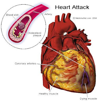
Coronary heart disease — All the organs and tissues in the body need a blood supply in order to function normally; blood carries oxygen and energy sources, especially sugar or glucose, that are required to meet the body's needs. It is the heart's job to pump oxygen-rich blood through the huge network of arteries that extend throughout the body, and this includes pumping blood into the vessels that supply the heart muscle itself. These vessels, called coronary arteries, lie on the outside of the heart muscle before entering the heart muscle itself (show figure 1).

In coronary heart disease (CHD), the coronary arteries become clogged with fatty deposits. The deposits, called plaques, cause a narrowing of the arteries through which the blood reaches the heart muscle (show figure 2). This can create an imbalance between the supply of blood (or oxygen) available to an area of heart muscle and the demand for blood (or oxygen) that is needed at a given moment. This causes a condition known as ischemia, sometimes felt by the patient as chest discomfort (angina pectoris).

Plaque rupture and clot formation — Sometimes a plaque in a coronary artery bursts or ruptures, leading to the formation of a blood clot that may partially or completely block the artery. A blockage of this type is likely to cause damage to the heart muscle due to a lack of blood and oxygen supply. If there is damage, an MI, or heart attack, has occurred.

The factors leading to the rupture of a plaque and the formation of a blood clot are not completely understood. There are certain characteristics related to the composition of a plaque that seem to make it vulnerable to rupture. Plaques that have an irregular appearance may contain less calcium and scar tissue and more lipids (fat) within their core, and may be more prone to rupture. Intense physical activity or psychologic stress immediately preceding MI may play a role because these activities affect the heart rate and blood pressure and could cause disruption of a plaque.

Some studies have shown that an emotional or stressful event occurs in a certain percentage of people in the days or hours before an MI. In addition, an increase in the incidence of MI has been noted in populations where a disaster has occurred. Because mental stress is known to cause increases in heart rate and blood pressure as well as other changes in the cardiovascular system, it has been suggested that, in some cases, mental stress can affect the stability of a coronary artery plaque and "trigger" an MI.

SYMPTOMS — The "typical" complaint of a person having an MI is persistent chest discomfort, resulting from ischemia involving an area of heart muscle. But there is wide variability in the symptoms a person can have when an MI is occurring. Some patients have no symptoms at all. Others, particularly women, older adults, diabetics, and individuals with a prior diagnosis of heart failure, may have other symptoms but not report chest discomfort. Still others have a combination of chest discomfort and other complaints. Some people having an MI will experience sudden death, also known as cardiac arrest.

Ischemic chest pain in angina and MI — Recall that the obstruction in blood flow that occurs in patients with CHD can cause an imbalance between the supply of oxygen-rich blood to an area of heart muscle and the demand for oxygen that exists at a point in time. This causes a relative lack of oxygen to the muscle, or ischemia, and may be felt by the patient as chest discomfort or angina pectoris. When the imbalance is eventually corrected during the anginal episode—for example, the person may rest, thereby decreasing the heart's oxygen demand—the discomfort slowly goes away and no damage has occurred.

The chest discomfort that occurs in the setting of an MI, caused by a blood clot that more seriously obstructs blood flow, can be similar to an episode of angina, because it also arises from ischemia to the heart. It may last longer (an episode of angina usually lasts more than two but less than 20 minutes), and may not be relieved by the measures that often relieve angina, such as rest or the use of nitroglycerin under the tongue.

Many different conditions can cause pain in the chest. The ischemic chest discomfort of an MI can range from mild to severe, and typically has the following characteristics:

* Felt as a pressure, constriction, tightness, or squeezing, versus a sharp or stabbing pain
* Is not limited to a small area, but rather spreads through the chest
* May radiate to other areas of the body, including the upper abdomen, shoulders, arms, neck and throat, or lower jaw and teeth
* Comes on gradually and lasts more than a few seconds
* Is usually not made worse by pressing on the area of the chest that is affected, by taking a deep breath or by a particular position; however, lying down may make it worse, while sitting up may produce some reduction in intensity
* Is not relieved by antacids or food

Other symptoms of MI — A number of other symptoms can occur in the setting of an MI. They may occur in a patient with or without chest discomfort. These include:

* Shortness of breath
* Nausea, vomiting, or belching
* Sweating
* Palpitations
* Lightheadedness
* Fatigue
* Fainting
* Cardiac arrest (sudden cardiac death)

DIAGNOSIS — The doctor uses a combination of tools to diagnose MI. The main tools are:

History and physical exam — The healthcare provider will gather information about any symptoms the patient may have experienced and analyze the person's risk of having CHD. Chest discomfort in someone with multiple risk factors for CHD raises the suspicion that the discomfort is cardiac in origin. (See "Patient information: Chest pain"). A complete physical exam may reveal signs of ischemia, such as abnormal heart sounds or cardiac rhythm.

Electrocardiogram — An electrocardiogram, or ECG, gives a picture of the flow of electrical activity that causes the heart to beat. Damaged or ischemic areas may (or may not) show an abnormal pattern. There are very specific patterns that are associated with an MI, and the ECG is usually the most important method for diagnosing an acute MI.

Blood tests — In the hours following a suspected MI, the blood can be monitored for substances (enzymes or proteins) released by damaged heart muscle. The presence of these substances in certain concentrations suggests that heart damage, or MI, has occurred.

TYPES OF MI — There are different types of MI, based partly on the area of the heart that is damaged and partly on the extent of damage as visualized on the ECG.

Location — Depending on which coronary arteries are obstructed, different areas of the heart can suffer damage during an MI. The areas most frequently affected include:

* Anterior MI
* Inferior MI
* Lateral MI
* Posterior MI
* Right ventricular MI

ST elevation MI (STEMI) versus non-ST elevation MI (NSTEMI) — The wave patterns on an ECG provide information about how the heart is functioning (show figure 3). In general, an ST elevation MI is caused by complete obstruction of a coronary artery, and causes damage that involves the full thickness of the heart muscle. In contrast, a non-ST elevation MI is caused by partial obstruction of a coronary artery, and causes damage that does not involve the full thickness of the heart wall. The treatment for these two types of MIs may differ.

TREATMENT — Acute MI refers to a heart attack that is in progress, as opposed to one that has occurred at some time in the past. Treatment decisions are based in part on whether the patient has had a STEMI or NSTEMI. Other factors, such as the patient's age, extent of CHD, and complications that have occurred also help direct treatment. A full discussion of treatments given during and immediately after is available separately. (See "Patient information: Heart attack treatment"). Recovery after heart attack is also discussed separately. (See "Patient information: Heart attack recovery").

COMPLICATIONS — Treatment of acute MI is more effective than in years past, and patients in general have a better expectation of a favorable outcome. Still, complications can occur, and include:

Ongoing ischemia — Early treatments may fail to halt the ischemic process and expanding areas of the heart may become damaged.

Abnormal heart rhythms — The damaged heart muscle can cause disturbances in the way the electrical impulses, which activate the muscle and cause the heart to beat, travel within the heart. An erratic heart beat and/or abnormal heart rhythm can result, some of which can lead to poor heart function or sudden cardiac death.

Poorly functioning left ventricle — If the heart's main pumping chamber, the left ventricle, is seriously damaged, its ability to function can be impaired. This can lead to a number of related complications (show figure 4).

* Heart failure — Heart failure refers to a condition in which the ventricle is stiff or weak, meaning that it has difficulty pumping blood in the normal way.

* Low blood pressure — Low blood pressure can occur, risking lack of adequate blood flow to the rest of the body, especially the brain.

* Cardiogenic shock — Cardiogenic shock can develop if the weakness of the heart is very severe and the amount of blood it pumps out very reduced; as a result, all the organs and tissues of the body suffer from lack of blood flow.

Left ventricular aneurysm — This refers to an area of left ventricle that has become thin and scarred and bulges outward, moving ineffectively during contraction. Left ventricular aneurysm can lead to a poorly functioning left ventricle, described above. If the wall is very thinned, the aneurysm may rupture, usually resulting in death.

Mechanical problems — An MI can cause the rupture of the muscles that hold the mitral valve in place; this valve, located between the left atrium and left ventricle, prevents blood from flowing back into the left atrium when the left ventricle contracts (show figure 4). If the valve is damaged, or insufficient, there is an abnormal flow between these two chambers, known as mitral regurgitation. Rupture of the membrane separating the left and right heart chambers can also occur, as can rupture of the wall of the ventricle.

These complications can be treated to a greater or lesser degree of success, depending upon a wide variety of factors, including early diagnosis, seriousness of the complication, and availability of appropriate interventions (for example, PTCA or cardiac surgery).

PREVENTION — Although MI cannot always be prevented, there are a number of good health practices that can reduce the chance that a person will have an MI.

Quitting smoking, controlling blood pressure and diabetes, and reducing blood cholesterol levels reduce a person's overall risk for developing CHD and MI. (See "Patient information: Smoking cessation" and see "Patient information: High blood pressure treatment in adults" and see "Patient information: High cholesterol and lipids (hyperlipidemia)").

In some patients, taking a low dose of aspirin every day is recommended as a way to reduce the risk of heart attack and stroke, but this should not be done without consulting with a healthcare provider. (See "Patient information: Aspirin and cardiovascular disease").

Finally, good routine medical care (going to the doctor for regular check-ups) may mean that early signs of CHD are detected when steps can be taken that may prevent a future MI.

Wednesday 17 June 2009

Percutaneous Transluminal Coronary Angioplasty / Stent


Introduction

More premature deaths are caused by coronary artery disease than by any other disease process in North America. Coronary artery disease is the narrowing or obstruction of the vessels that supply blood and oxygen to the heart muscle. This narrowing is caused by fatty deposits (plaque) on the walls of the arteries.

Occluded LAD Coronary Artery These fatty deposits gradually build up and can cause a marked reduction of blood and oxygen to the heart. If the blood flow is significantly reduced, some form of medical treatment becomes necessary. The picture on the right shows a diseased LAD artery.

The Heart - Coronary Arteries


One of the most common non-surgical treatment for opening obstructed coronary arteries is Percutanueous Transluminal Coronary Angioplasty (PTCA). The name itself says a lot about the procedure:

Percutaneous means access to the blood vessel is made through the skin

Transluminal means the procedure is performed within the blood vessel

Coronary specifies that the coronary artery is being treated

Angioplasty means "to reshape" the blood vessel (with balloon inflation)Also referred to as "balloon treatment" because special balloons are used to open up obstructed arteries, illustrated on the left, this procedure sometimes also involves the use of devices known as "stents" to help keep the arteries open.

Balloon Catheter Opening ArteryThe illustration on the right shows how a balloon catheter works to open an occluded artery.


Below, on the left, is an illustration of a special catheter being used to install a stent device. If agreed upon in advance by you and your physician, the diagnostic cardiac catheterization procedure and PTCA are done together. This is an added convenience for you as it shortens your hospital stay by one day.Balloon Catheter Opening Artery


LAD after PTCA/stentThe picture on the right shows the diseased LAD pictured above after it has undergone a successful PTCA procedure in which a stent was installed.


Following is a description of the PTCA procedure. We hope to answer some of the questions you might have. However, it is important for you to realize that everyone's situation may be different, and what you actually experience may vary slightly from what you read on the following pages. If you have any questions or doubts, please contact us or your physician.


PTCA/Stent Procedure
Pre-Admission and EvaluationOccluded Artery

You will first undergo a thorough evaluation by your physician. Any questions you still have before your procedure begins can be answered at this time. You may be permitted clear liquids in the morning of the procedure. However, this should be cleared with your physician first . Otherwise, food and beverages may be withheld after midnight. If you are a diabetic, you will be given special instructions. Your groin area will be washed and shaved in preparation for the PTCA/Stent.


Angioplasty Procedure

If family or friends are with you, they will wait in the waiting room near the catheterization laboratory. The length of the procedure depends on the complexity of each individual's situation, but in general, the duration is between 1 and 2 hours.

An intravenous line will be started in your arm this morning. You will receive various medications in the angioplasty laboratory though this line. To help you relax, you will be given medication prior to leaving for the lab. You will remain awake, but slightly drowsy.

You will be placed on an x-ray table upon your arrival in the lab. It is the same type of room in which you had your cardiac catheterization. All personnel in the lab will be wearing surgical attire. You will be covered by sterile sheets, and so will some of the equipment. Your groin (arm) will be cleansed with an antiseptic (might be cold) and then numbed with an anesthetic. You will feel the sting of the needle, but then your groin (arm) will feel quite numb. Heart monitoring equipment will be placed on your arms and legs, and you may be given oxygen to breathe. You will be given certain medications through the intravenous line, and periodically medication will be given to relax you and decrease any restlessness. Remember, you must still be able to talk and follow directions.


The NIR stent shown in the delivery
state (above) and in the expanded
installed state (below).
The angioplasty catheter (balloon-tipped) is inserted at the numb area, and advanced to your heart, using x-ray to guide it. When the balloon is inflated at the point of the blockage, you may feel chest pressure, or discomfort, and this is normal. It will subside when the balloon is deflated. You may also feel your heart thump or skip, feel flushed, or have a headache. All these sensations are normal. You will be asked at times to hold your breath for a few seconds. You may also be asked to cough.

After the procedure, you will be moved to a recovery area for a short time, and then taken to your room where your heart can be monitored. Nurses will closely monitor your vital signs and general well being. They will also frequently check the groin area and dressing. A small, flexible catheter is routinely left in the groin for 4-6 hours unless a percutaneous suture is used to close the hole. You will be required to remain in bed and keep your leg immobilized.

You will be able to eat as soon as you wish after the procedure.

Recuperation

The catheter or sheath will be removed approximately 4-6 hours after theprocedure is over. This waiting period is crucial as the physician uses blood -thinning agents to implant the stent, thus the sheaths cannot be removed until the blood thinning reverted back to normal and firm presssure applied for about 20 minutes. Then a pressure bandage is applied and a small sandbag placed over it. This is to assure proper healing of the artery. Pain medication is available to you every few hours after the procedure. Please let your nurse know of any pain or discomfort you may feel at any time. The rest of the day is basically for rest, recuperation, and a gradual return to your activities.


Discharge

Your doctor will see you the morning of discharge. Your doctor's nurse will go over medications, activities, and follow-up care. You will receive prescriptions for medicines you will need, and will usually undergo a low-level stress test very shortly after your discharge.

POSSIBLE COMPLICATIONS AND RISK

No invasive procedure occurs without a certain amount of potential risk and complications. You are followed closely by your doctor and nurses, precisely for the reason that if any of these complications arise, corrective action can be taken immediately. The incidence of complications is low, but nonetheless, you need to be aware what they can be:


1. Severe bruising/bleeding into the groin area of the procedure leg.
2. Changes in your heart rhythm.
3. Allergic reaction to the contrast or dye or to other medications used.
4. A tear in the lining of the artery which is being dilated.
5. Possibility of a heart attack during or following inflation of the balloon or stent implantation.. (you are given strong blood thinners to minimize this risk.)
6. Possibility of a blood clot in the artery in which the catheter is inserted. (If this occurs, surgical removal may be required.)

Follow-up Care

Periodic follow-up with your personal physician is quite important. It is important for you to realize that by angioplasty (PTCA), your immediate problem has been taken care of, but it does not cure coronary artery disease. In some patients, re -narrowing (Restenosis) of the artery may occur over the ensuing 3-6 months. If this happens, your original symptoms may return, or your stress test will be abnormal.

If you have chest discomfort, stop your activity, sit or lie down, and take nitroglycerine, as instructed. If the chest discomfort does not go away after 3 nitrolycerin tablets in a 15 minute period, DO NOT DELAY SEEKING MEDICAL ATTENTION. Either call your doctor, or go to an emergency room.